

 Login
Login35 yo male, brain tumor, exact site not specified
Immunos
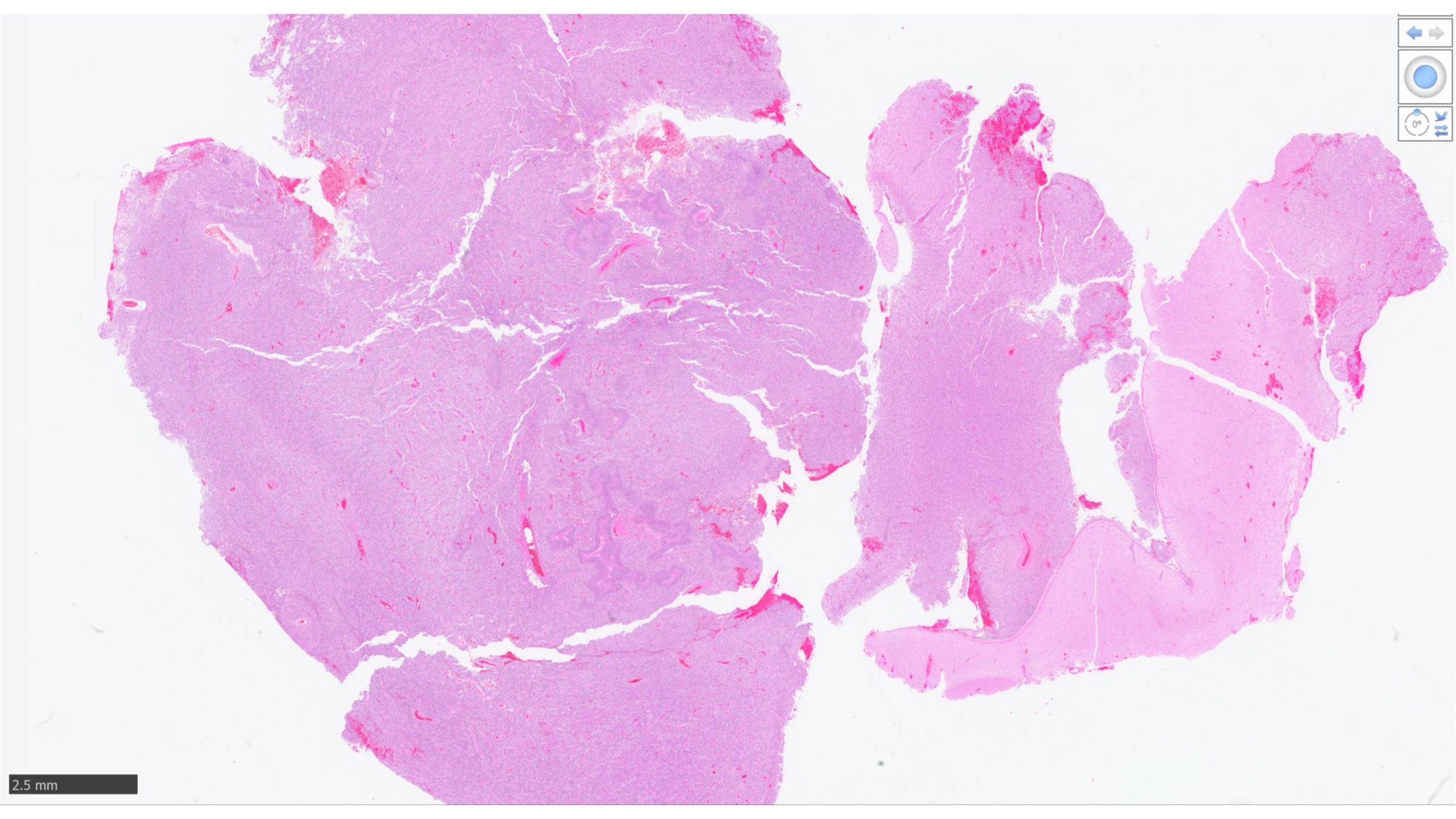
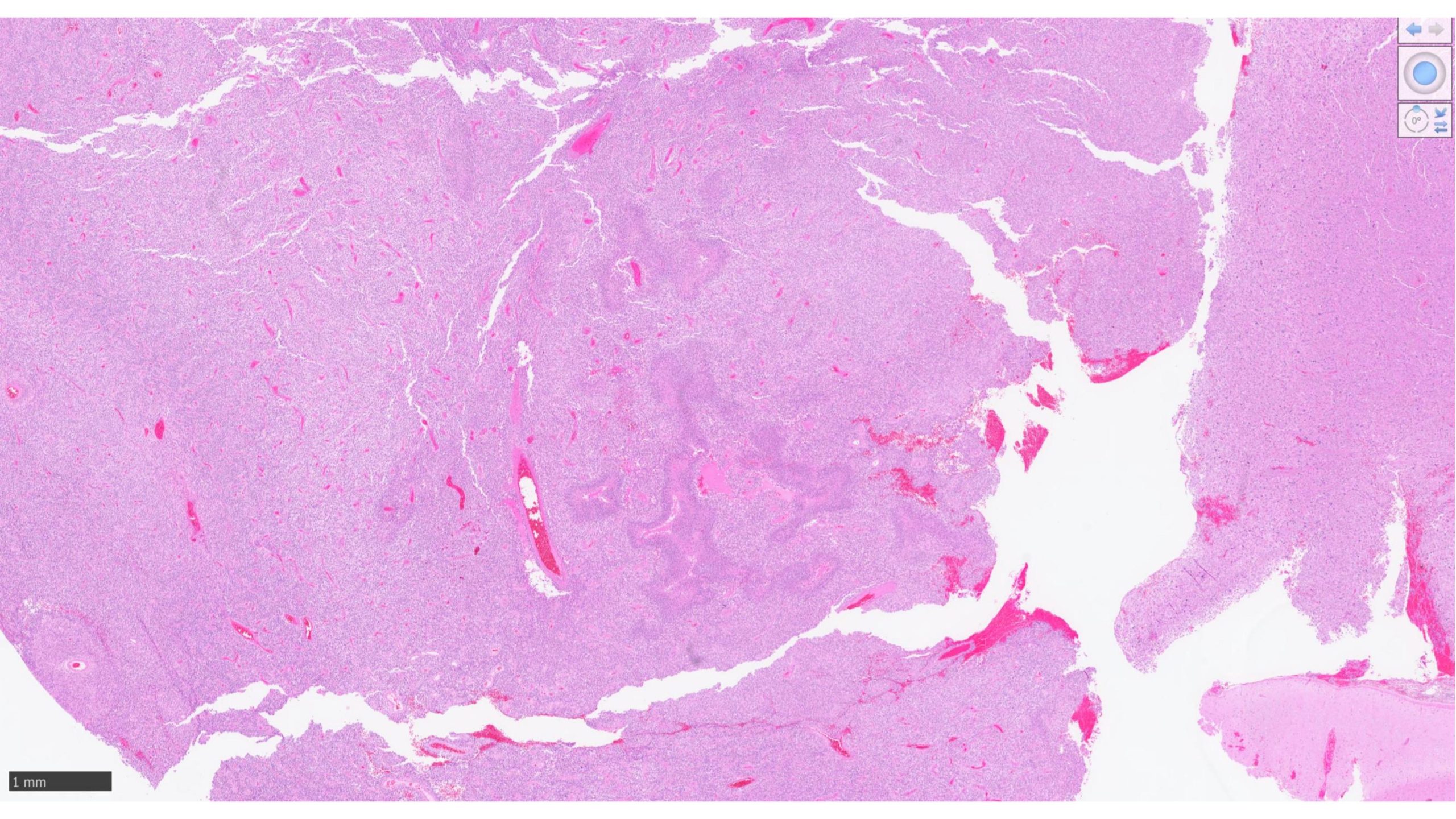
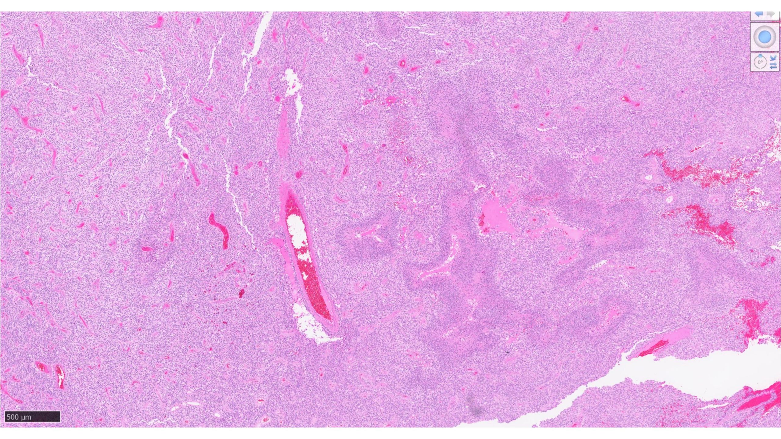
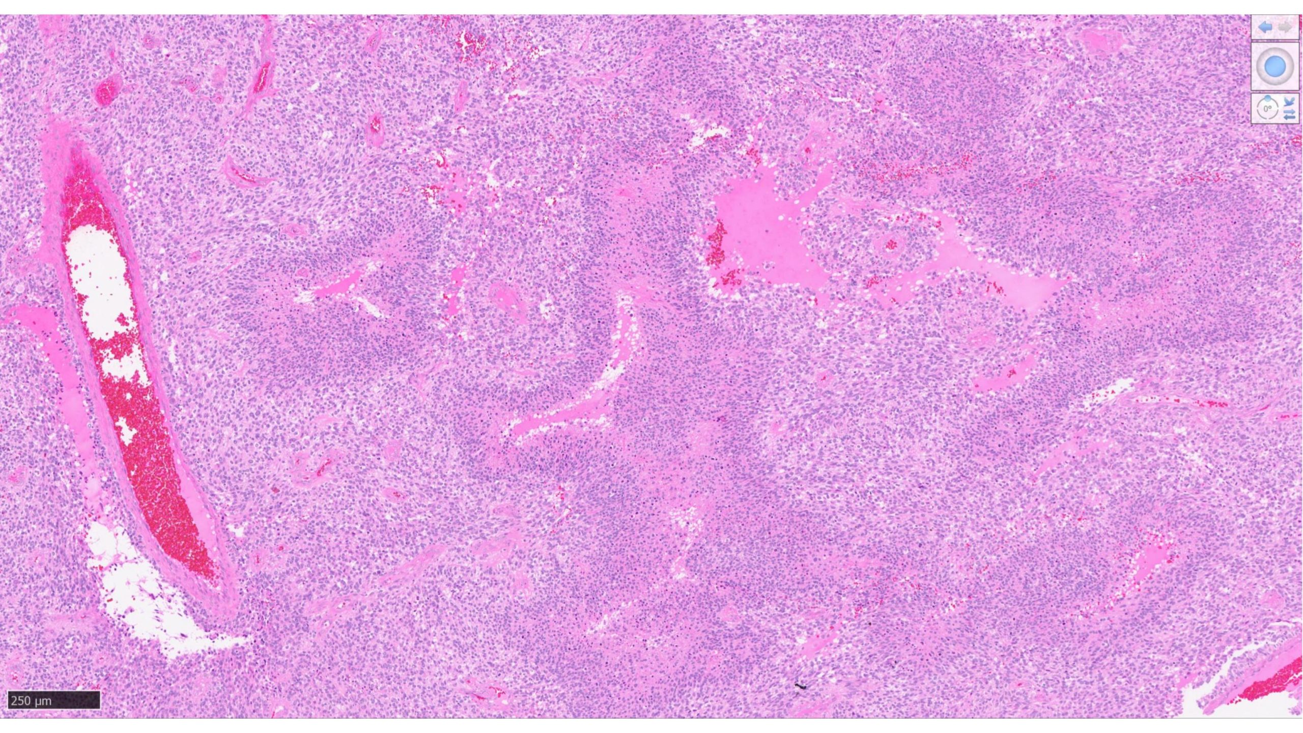
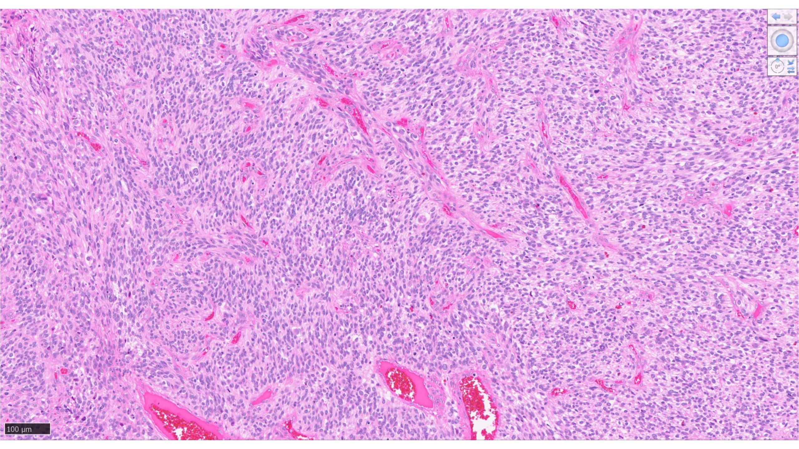
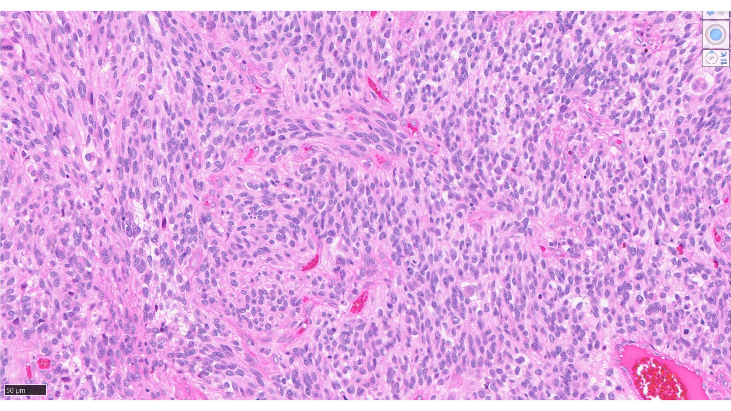
Sections show a highly cellular glial neoplasm with cellular atypia including bizarre cells, frequent mitoses, vascular proliferation and palisading necrosis. Immunohistochemistry stains with appropriate controls were performed on block B5. Tumor cells are GFAP positive, Olig2 shows a mixed pattern of staining with positive and negative cells. ATRX is lost in tumor cells. P53 is diffusely positive( presumed mutant pattern). IDH1 is negative. Ki-67 is elevated (more than 30% in areas). The morphology of this neoplasm is that of a high-grade glioma. Molecular studies are being performed and an integrated diagnosis will follow in an addendum.
#
 Programação completa
 Palestrantes confirmados
 Comissões
 Trabalhos e seminários
 Localização do evento
 Valores